Business opportunity
Scientists at the Inst. of Clin. Med., Univ. of Oslo, Akershus Univ. Hospital, and Faculty of Med., Univ. of Geneva are developing a tool for fully automated segmentation (creation of 3D models) and labelling of all blood vessels in a few seconds. The AI-based tool can be applied to any abdominal computer tomography (CT) dataset.
The user of the tool will typically be a cancer surgeon who uploads the input medical CT images from an individual patient to a secure cloud/server, runs the algorithm, and creates a 3D model output. The surgeon gets a precise map of the patient-spesific anatomy allowing correct interpretation of the individual anatomy before and during surgery. With this navigation tool, surgeons can perform many cancer surgery procedures with greater confidence. As a result, this will lead to faster and safer surgical procedures which ultimately will have a positive health and economic impact.
A major part of cancer surgery is based on extensive lymphadenectomy, removing as many lymph nodes as possible with the hope to reduce the chances of relapse. There is a positive correlation between the number of lymph nodes removed and increased survival rate in cancer surgery. To offer this type of surgical treatments, it is crucial for the surgeon to have detailed knowledge of the patient-specific vascular structures beforehand, because the pattern of the lymph node drainage follows the pattern of blood vessels. Unfortunately, surgeons are presently limited to the information they get from 2D pictures of CT scans which are usually less detailed than desired. We offer a solution to this problem through the creation of detailed patient-specific 3D models giving a 360 degrees view of the region of interest.
Inven2 seeks partners for co-development and can offer exclusive access to the software and know-how. We are interested to validate the technology applicability together with a user partner.
Technology
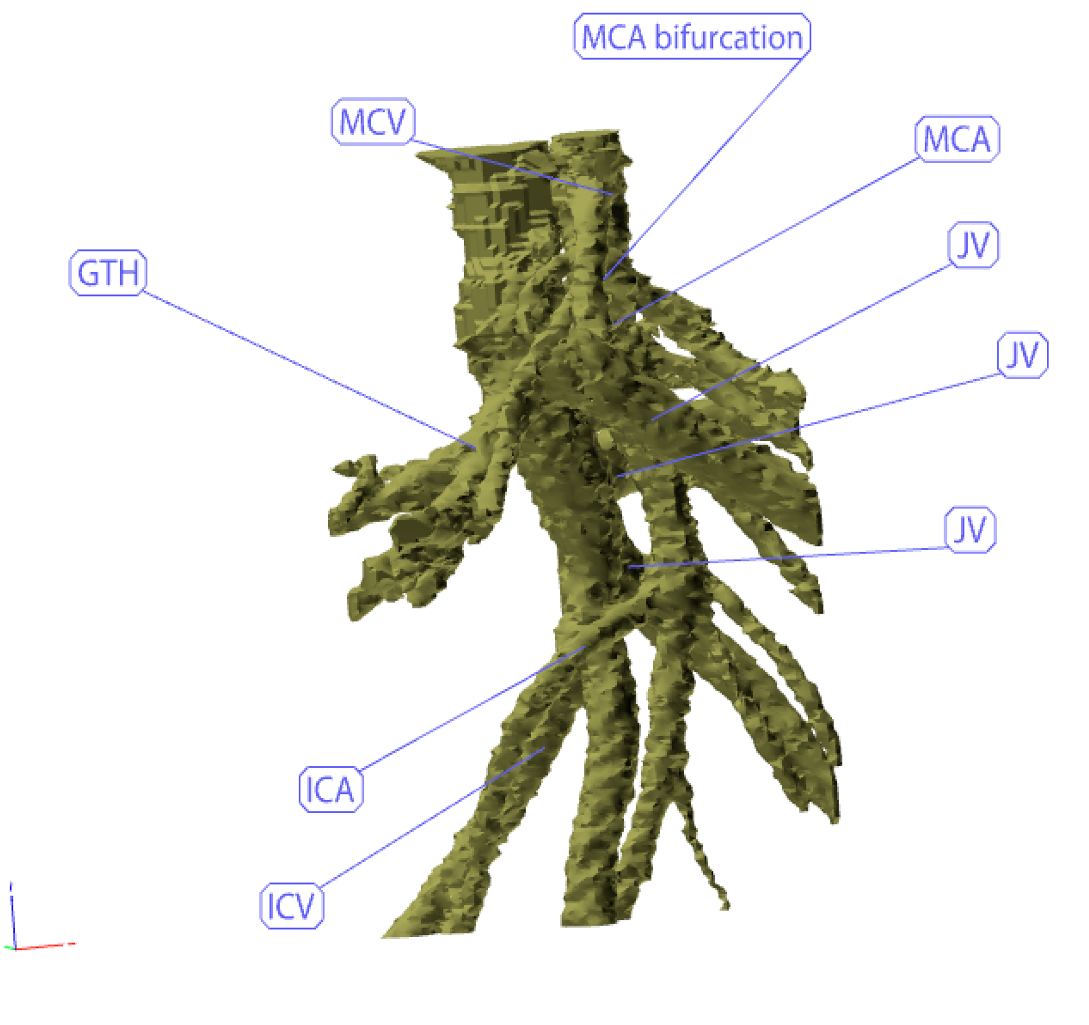
We have data from more than 500 patients containing CT images with corresponding clinically-validated segmented and annotated models. This data material is now used to develop the automatic tool through neural network training. Every vessel branch in the training dataset is annotated (labelled) with its corresponding anatomical name. This information will give the surgeon a very detailed map of the anatomy during surgery.
Advantages
The tool will provide detailed and individual vascular information (even on very small vessels) to cancer surgeons in an automatized and scalable way.